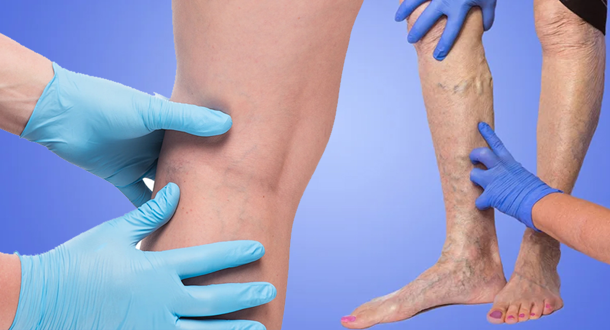
Venous Hypertension

What is venous hypertension?
Venous diseases are one of the most predominant diseases in the medical profession. It is claimed that 50% of the world population gets venous problems some time in their lives. Like arterial hypertension which we all have heard of, venous hypertension is the root cause of all venous diseases.
What is the anatomy of the venous system in the legs?
These comprise the :
- Superficial venous system in the legs
- Deep venous system in the legs
- Perforating veins which connect the superficial to the deep venous systems.
Content Reviewed by - Dr. Jaisom Chopra
Causes
Why does it happen?
There are two basic causes of venous hypertension :
- Blockage of the veins
- Incompetence of the valves within the veins.
| Superficial veins | Deep veins | |
| Blockage | phlebitis | Deep vein thrombosis (DVT) |
| Incompetence | Varicose veins | Chronic venous insufficiency (CVI) |
How do they present?
- Dull Ache
- Swelling
- Heaviness
- Night cramps
- Leg tiredness
- Restless legs
18% are continuous symptoms while 50% are episodic.
Episodic pain due to varicose veins may be hormonal. Half the pregnant women with varicose veins have pain in the legs and 17% are unable to remain straight for 1-2 hours at a stretch due to the severity of pain.
Walking and leg elevation bring relief along with compression stockings. Warmth aggravates the pain and cold relieves it.
Leg pain may be present much before varicose veins present.
Who is more prone to venous hypertension?
- Age - incidence increases with age
- DVT - Causes dilatation of the vein due to blockage leading to increased venous pressure.
- Sedentary life - By this, the pump action of the calf muscles is minimized causing stagnation of blood and thus venous hypertension.
- Obese women - it is commoner.
- Prolonged standing jobs - lead to increased venous pressure.
- Smoking - Higher incidence in male smokers.
- Pregnancy - is an important causative factor.
How common is it?
- 2 - 5% of all adult population suffers from CVI
- Varicose veins present in 7 – 60% of the adult population and 40% of them have venous reflux.
- Venous stasis is seen in 3% of the population
- Hospital admission for CVI is 92/100,000
- The incidence of CVI is higher in industrialized nations than in developing countries due to differences in lifestyle and activity.
What is the sequence of events in venous hypertension?
- Venous hypertension increases the morbidity and disability of patients with venous disease. If no treatment is done the condition only worsens due to the progression of a disease. The skin eventually breaks down and venous ulcers result. These ulcers are recurrent. This is preceded by thickening of the skin or lipodermatosclerosis.
- Chronic non-healing ulcers increase the morbidity and only heal if the reflux is totally eliminated.
- Tissue atrophy and colour changes are not reversible.
Why are there colour changes and ulcers around the ankle?
The dilated veins stagnate the return of blood causing the pooling of cells mainly WBC which release proteolytic enzymes which damage the lining of the thin capillary veins. This allows the fibrinogen to pass out into the tissue and form a wall which prevents tissue around to starve of oxygen and thus they break down. The colour changes are due to the deposit of the deposition of hemosiderin from the RBC into the tissue.
How do you diagnose venous hypertension?
Misdiagnosing venous occlusion may be lethal but venous insufficiency is slowly progressive leading to reduced quality of life.
Blood for D-dimer is always elevated due to recurrent venous thrombosis but it reduces the usefulness of detecting DVT.
Colour Doppler study
It is the investigation of choice. It tells us about any dilatation, blockage and can predict venous hypertension. It is non-invasive.
Link showing venous reflux https://www.youtube.com/watch?v=P15g-Oewbys
Venography
MR Venography is most sensitive and specific for venous disease of the legs and pelvis. It can detect unsuspected non-vascular causes of leg pain and edema.
Venous Plethysmography
here infra-red light assesses the capillary filling during exercise. An increased capillary filling is indicative of venous reflux and thus incompetent veins.
Physiologic venous function tests
These predict the severity of venous insufficiency. The parameters used are:
- a) Venous Refilling time (VRT)
- b) Maximum Venous Overflow (MVO)
- c) Calf Muscle pump Ejection Fraction (CMEF)a) Venous Filling Time (VRT)
The patient is made to lie down and the calf muscles are emptied of blood using a calf muscle pump. This is done as thoroughly as possible. Now the patient is made to sit and the filling of the veins in the calf occurs by the arterial blood and takes over 2 minutes or 120 secs. If this filling takes place between 40 – 120 secs it means there is mild venous reflux across early leaky valves. If this venous filling takes place between 20 – 40 secs then there is sufficient venous insufficiency due to a failure of the valves in the superficial and the perforating venous systems. These patients have leg cramps, restless legs, burning leg pain, and premature leg fatigue. Should the VRT be less than 20 secs? It shows high venous reflux from the superficial, perforating and the deep veins. These patients are symptomatic. Patients with VRT less than 10 seconds mostly have venous ulcers.
Maximum venous overflow (MVO)
This detects the obstruction to the venous outflow in the leg irrespective of the cause. The results show the speed with which blood flows out of a maximally congested leg when an occluding tight tourniquet is suddenly removed. It detects occlusions in the calf veins, iliac veins, and IVC where colour Doppler and venography are insensitive. The test is not very sensitive with partial obstruction and reflux-induced venous insufficiency. A normal MVO does not rule out DVT categorically.
Muscle Pump Ejection Fraction (MPEF)
It detects the failure of the calf muscle pump to expel blood from the lower leg. The patient stands on the toes 10 – 20 times or dorsiflexes the ankle. The change in blood volume within the calf muscles is recorded as the calf muscle is pumped. Normally in 10 – 20 ankle dorsiflexions the calf muscles empty. In patients with calf muscle pump failure or severe proximal obstruction or severe deep venous insufficiency, there is little effect on the calf muscles.
What happens if I do not treat venous hypertension?
- There is an increased lifetime risk of DVT and PE.
- Amputation is needed in 1.2% of the people
- Overall mortality is 1.6%
- 50% of patients with untreated varicose veins develop thrombophlebitis at some time.
- 45% of patients with superficial phlebitis have unrecognized DVT.
- The risk of DVT is 3 times higher in Varicose veins patients.
- Venous hypertension patients on bed-rest and allied illnesses are prone to DVT.
- Phlebitis develops in 60% venous hypertension patients and 50% of these patients develop DVT.
- The incidence of PE is higher in venous hypertension patients developing DVT (50%) and the death rate is also 1 in 3 from PE.
- Venous Hypertension patients die from hemorrhage due to bleeding varices.
Content Reviewed by - Dr. Jaisom Chopra
Treatment
What are the approaches to the treatment of venous hypertension?
- Venous hypertension is not uncommon.
- Treatment is directed to relieve symptoms and where possible the cause.
- No oral medicine has been found which can cure venous hypertension.
- Surgical or endo-venous therapy is for ulcers not responding to medical treatment.
- Graded compression stockings are the cornerstone of the modern treatment of venous hypertension.
- The main aim of treatment is to remove, if possible, major reflux pathways.
- Valvuloplasty is occasionally helpful but the incidence of post-operative DVT is high.
- A refluxing vein is no longer needed and can be ablated without side effects.
- Antibiotics are not useful in venous ulcers unless infected.
What is the role of compression stockings?
- These provide 30 – 40 mmHg or 40 – 50 mmHg compression at the ankle and reducing compression as we move upward. This helps restore venous pressure to normal and improve venous flow in patients with severe venous incompetence.
- The graded compression is vital as non-graded ones give a tourniquet effect and worsen venous reflux.
- The anti-embolic stocking is useless and neither improve venous return nor prevent venous thromboembolism.
- Leg elevation is helpful in promoting venous return due to gravity and reduces edema. On sitting legs should be above the level of the thighs and on lying above the level of the heart.
- Four layer compression dressings are the main-stay in venous ulcer cases. They contain calamine lotion, glycerine, zinc oxide, and gelatin.
What are the various treatments possible?
- The patient must be instructed that leg elevation and compression stockings are his life-line and he must not stand or sit for long at one site with legs dangling. They must walk and do calf exercises at regular intervals.
- Thrombolysis using TPA or thrombectomy have poor results with a very high recurrence rate.
- Saphenous vein bypass for iliofemoral venous occlusion shows poor results with failure rates about 30%.
- Surgery for CVI includes valvuloplasty and allografts or cadaveric vein transplants.
- Congenital absence of valves have valvuloplasty with perforator ligation but the success rate is 70% success after 5 years.
What is the latest surgical therapy available?
- The latest treatment all over the world is Radio-Frequency Ablation (RFA) or Laser therapy (EVLT) along with Foam Sclerotherapy.
- Both RFA and EVLT are endovenous ablation techniques. In both techniques, energy is delivered to the lining of the diseased vein which destroys it and causes it to fibrosis. They have provided excellent results over 10 years of study.
- Sclerotherapy uses a sclerosing agent (STD) which destroys the lining of the diseased refluxing veins which fibrosis.
- Subfascial Endoscopic Perforator Surgery (SEPS) – has been used to treat CVI. The perforating veins are ligated endoscopically. After the treatment, the average healing time for venous ulcers is 42 days with a recurrence of 3%. Ulcers with this technique heal 3 times faster.
How are bleeding veins treated?
Immediate sclerotherapy followed by compression and limb elevation is the treatment of choice.
Are there any complications of surgery?
- Sclerotherapy - allergic reaction to sclerosant; Skin necrosis after extravasation of the agent.
- RFA or EVLT - Skin burns; Thermal injury to pre-venous tissue; injury to deep tissue.
What activity is recommended?
- Regular exercise is advisable.
- Prolonged standing or sitting is not well tolerated.
- Walking, running, cycling and swimming is excellent if the muscle pump is intact.
- Patients with obstructed venous flow have increased pain and swelling on exercise.
- Patient with muscle pump failure has markedly reduced exercise tolerance due to leg fatigue.
Can venous problems be prevented?
At the first suggestion of venous disease :
- Avoid prolonged standing
- Using graded compression below knee stockings with a pressure of 30 – 40 mmHg.
Content Reviewed by - Dr. Jaisom Chopra


