


Aneurysm (Aneurysmal Dilatation)
What is Aneurysm
An aneurysm is a blood-filled balloon in the wall of a blood vessel. They could involve any blood vessel in the body.
An aneurysm dilates and could rupture leading to massive blood loss followed by marked hypovolemia and finally death. The blood vessel could be thin as a result of a hereditary cause or acquired.
Aneurysm could be the site for clot formation and embolization.
Classification of Aneurysm
Aneurysms could be classified by type, morphology, and location.
True or False type of aneurysms
True aneurysm
involve all 3 layers of the arterial wall (intima, media, and adventitia). These are seen in atherosclerosis, syphilis and congenital aneurysms. The ventricular aneurysms that follow transmural myocardial infarction involve all layers of the wall of the heart and are called true aneurysm.
False aneurysms
are also called pseudo-aneurysms is a collection of blood leaking out of the artery wall and confined to the vessel by the surrounding tissue. This blood-filled cavity will eventually thrombose and seal the leak or rupture out of the surrounding tissue. Pseudoaneurysms are caused by trauma that punctures the artery wall like knife and bullet wounds or percutaneous surgical procedures like coronary angiography or arterial grafting or using an artery for injection.
Morphological classification
This is according to the shape and size and may be saccular or fusiform.
Saccular aneurysms
They are rounded in shape and involve only a portion of the vessel wall. They are 5-20 cm in diameter and are filled partially or fully by thrombus.
Fusiform aneurysms
are spindle-shaped and vary both in diameter and length. Their diameter can extend to 20 cm (8 inches). They involve large sections of the transverse and descending aorta and less frequently iliac arteries.
Classification according to the location
They could be:
Arterial and venous aneurysms
- Heart coronary artery aneurysms, ventricular aneurysms, aneurysm of sinus of Valsalva, and aneurysms following cardiac surgery
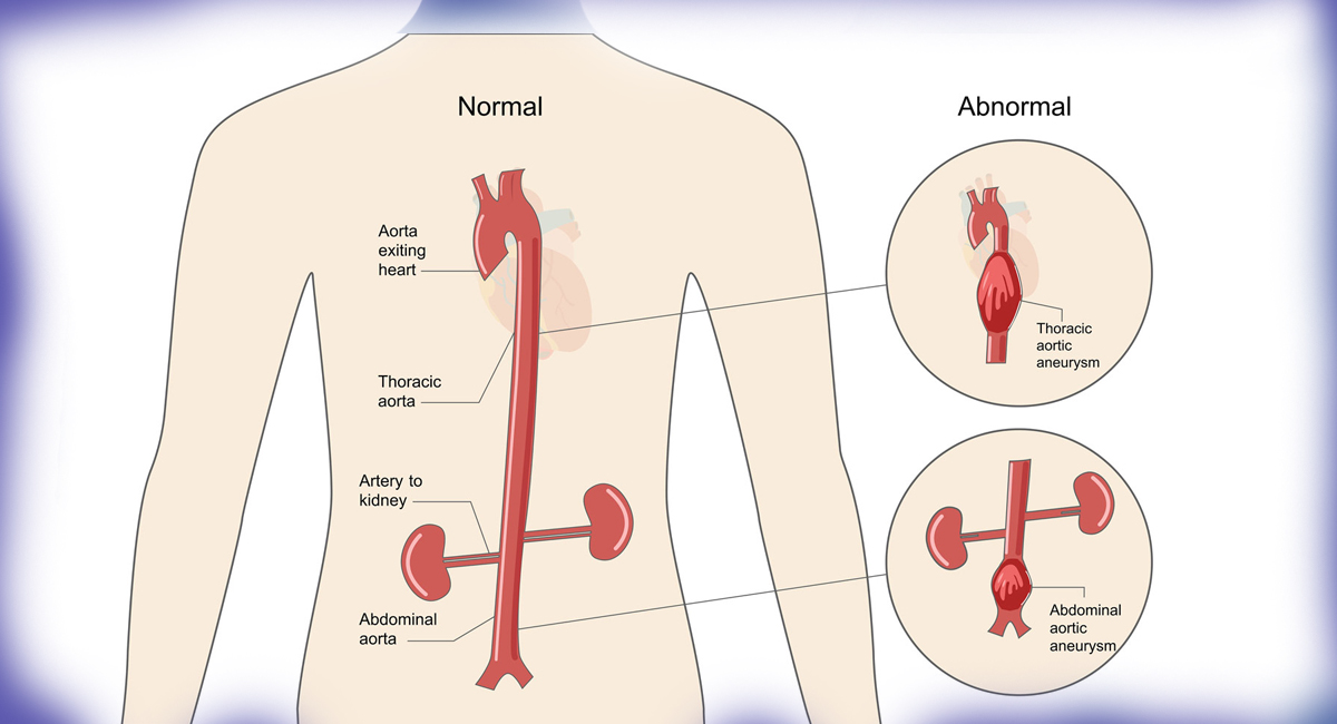
- Aorta - thoracic or abdominal aortic aneurysms
- Brain - berry aneurysms, cerebral aneurysms, and Charcot-Bouchard aneurysms
- Legs - popliteal artery aneurysms
- Kidneys - renal artery aneurysms
- Capillaries - capillary aneurysms
Cerebral aneurysms occur in the anterior cerebral artery which is part of the circle of Willis. Its rupture can cause severe stroke leading death. The second commonest site is the internal cerebral artery.
Content Reviewed by - Dr. Jaisom Chopra
Facts
Aneurysm facts
- An aneurysm occurs when part of the blood vessel or cardiac chamber swells.
- The aneurysm can occur in any part of the body though they often occur in the wall of the aorta.
- The two main types of aneurysms are aortic and cerebral.
- Aneurysms are more common in men and people over 65 years.
- Symptoms depend on the size of the aneurysm and how rapidly it is growing and its location.
- A large cerebral aneurysm can lead to numbness of the face or problems with the eyes.
- Cerebral aneurysms generally form at the base of the brain.
- 1 in 4 aortic aneurysms is in the thorax.
- The aim of the treatment of aortic aneurysms is to prevent its rupture.
- A ruptured brain aneurysm can be treated by surgical clipping or endovascular coiling.
Symptoms
Signs and symptoms of Aneurysm
Aneurysmal symptoms vary from asymptomatic to hypovolemic shock. They vary according to the site:
Cerebral aneurysm
The symptoms depend upon the pressure of the aneurysm on the brain structures and if it has ruptured or not.
The symptoms in a case where the aneurysm has not ruptured are:
- Fatigue
- Nausea and vomiting
- Seizures
- Loss of consciousness
- Loss of perception
- Loss of balance
- Speech problems
- Double vision
In case of a ruptured aneurysm (subarachnoid hemorrhage) the symptoms are:
- Severe headache
- Loss of vision
- Double vision
- Neck pain or stiffness
- Pain above or behind the eyes
Abdominal Aneurysm
- Central back pain
- Throbbing sensation in the abdomen
- Edema legs and deep vein thrombosis
- Vomiting
- Lower limb ischemia
Thoracic aortic aneurysms
- Coughing
- Loss of voice
- Breathing difficulty
- Problems swallowing
Renal (kidney) Aneurysms
- Flank pain
- Hypertension
- Haematuria
- Hypovolemic shock
Content Reviewed by - Dr. Jaisom Chopra
Causes
Causes of Aneurysm?
Brain (cerebral) aneurysm Potential causes:
- Weakness in the arterial wall (usually since birth)
- Hypertension.
- Atherosclerosis (plaques of cholesterol, platelets, fibrin formed in the arterial wall).
- Age at menopause influences cerebral aneurysm risk - early menopause increases the risk.
- Most aneurysms develop at the bifurcating folk of the artery as it is weaker. They mostly form at the base of the brain but could be present anywhere.
Abdominal aortic aneurysms (AAA) causes:
- Atherosclerosis.
- Smoking is a major risk factor as smoking contributes to atherosclerosis, hypertension, and acceleration of aneurysmal growth. Women smokers are 4 times more likely to develop AAA.
- Poorly controlled hypertension.
- Vasculitis (infection in the aorta) is mostly familial.
- Cocaine users had 4 times more risk of coronary artery aneurysms.
- Gene linked - the gene linked with cancer and cardiovascular development is also linked to AAA.
- Traumatic injury - caused by vehicle accident of a bad fall.
Thoracic Aortic aneurysm causes:
These are the same for AAA with the addition of:
- Marfan syndrome - a disorder of connective tissue.
- A previous aortic injury like aortic dissection.
Content Reviewed by - Dr. Jaisom Chopra
Risk Factors
Risk factors of Aneurysm
- Diabetes
- Obesity
- Hypertension
- Tobacco abuse
- Alcoholism
- High cholesterol
- Copper deficiency
- Increasing age
- Tertiary syphilis infection
Specific infective causes associated with aneurysm include:
- Advanced syphilis infection is responsible for syphilitic aortitis and aortic aneurysm.
- Tuberculosis causing Rasmussen's aneurysms.
- Brain infection causing an infectious intracranial aneurysm.
Aneurysms associated with genetic factors are:
- Berry aneurysm of the anterior communicating artery of the circle of Willis associated with autosomal dominant polycystic kidney disease.
- Familial thoracic aortic aneurysms.
- Cirsoid aneurysms were secondary to congenital Arteriovenous Malformations.
Pathophysiology
Atherosclerosis
weakens the blood vessel wall while repeated trauma of blood flowing through the vessels contributes to the degeneration of the vessel wall. hypertensive injury compounds the degeneration and accelerates the expansion of the aneurysm. This expansion of the wall leads to increased tension. The pressure of the blood within the expanding aneurysm injures the blood vessels supplying the wall of the artery further weakening the arterial wall. Without treatment, these aneurysms progress to rupture.
Infection
Mycotic aneurysm is due to a bacterial infection that involves the arterial wall. The common locations are the abdomen, thigh arm, and neck. The complications are rupture or sepsis. Less than 3% of the abdominal aortic aneurysms are mycotic.
Syphilis
In its 3rd stage can manifest as aneurysm of the aorta which is due to loss of the vasa vasorum in the tunica adventitia.
Copper deficiency
Though this is very rare it is due to reduced activity of the lysyl oxidase enzyme affecting elastin which is a key component in the arterial wall. It leads to thinning of the arterial wall and thus aneurysm formation.
Content Reviewed by - Dr. Jaisom Chopra
Diagnosis of Aneurysm
Aortic aneurysm diagnosis
Mostly they are an incidental finding when a patient is being examined for another ailment. If suspected the investigations ordered are:
- Abdominal ultrasound- It is a non-invasive test where it is possible to know the presence of the aneurysm and the size.
- CT Scan It is possible to produce a three-dimensional picture of the aneurysm.
- MRI Scan - magnets and radio waves produce a 2 dimensional and 3-dimensional pictures of the target area.
- Screening program - for the asymptomatic over 60 years of regular smokers or a family history of AAA.
Cerebral aneurysms diagnosis
This is suspected in patients with sudden severe headache, pain behind the eyes, vision changes, paralysis of one side of the face should have the following tests:
- CT Scan - create a 3-dimensional picture of the target area. Multidetector computerized tomography (MDCT) angiography is highly accurate in detecting intracranial aneurysms.
- Cerebrospinal fluid test - in cases of subarachnoid hemorrhage there will be RBCs in the CSF. This is indicated for patients who have symptoms of the ruptured aneurysm but CT was negative. It is a lumbar puncture or spinal tap.
- MRI Scan - is better than CT in detecting a ruptured aneurysm.
- CT Angiogram - Shows a rupture of the aneurysm. It is only used when others have failed as it is an invasive procedure.
Content Reviewed by - Dr. Jaisom Chopra
Aneurysm Treatment
There are currently two options for brain aneurysms - surgical clipping and endovascular coiling.
- In surgical clipping, a craniotomy is done to expose the aneurysm the base is closed by a clip.
- In endovascular coiling, a catheter is passed from the groin through the aorta to the brain arteries and the aneurysm is entered and platinum coils are introduced. They introduce a clotting reaction that fills the dome and prevents rupture.
One-third of brain aneurysms rupture. Size is not a significant risk factor.
The following measures help relieve symptoms and manage complications:
- Painkillers for headaches
- Calcium channel blockers - prevent calcium from entering cells of the blood vessels. They reduce the amount of narrowing and widening of the blood vessels, a complication of the ruptured aneurysm.
- A vasopressor - rises the blood pressure and widens the narrowed blood vessels. The aim is to prevent a stroke.
- Anti-seizure drugs - occur after rupturing of the aneurysm -phenytoin.
- Ventricular catheter - prevents hydrocephalus by draining collected fluid in the brain spaces into an external bag. A shunt may be needed. This is a flexible rubber tube with a valve starting in the brain and ending in the abdominal cavity draining the excess fluid into the large abdominal cavity.
- Rehabilitation therapy - there may be brain damage causing body and speech impairment. This therapy helps the patient relearn vital skills.
- Treatment of unruptured cerebral artery aneurysm.
It may be sealed off by surgical clipping or endovascular coiling. The risks are almost equal and may be higher than the potential benefits. The decision making is helped by:
- A site of the aneurysm
- Size of the aneurysm
- Patient's age
- Patients general condition
- A family history of ruptured aneurysm
- Other congenital conditions which increase the risk of aneurysm rupturing.
Aortic and peripheral aneurysms
The treatment is dependent on two choices:
- 1. Wait and watch
- 2. Surgery
How big is the aneurysm and how fast it is growing?
- If the aneurysm is small (4cm or less) and asymptomatic. Wait and watch and perform ultrasound every 6 - 12 months. The patient is informed of the signs and symptoms of dissection or rupture.
- Statins reduce the growth rate of small aneurysms.
- Medium sized aneurysms (4 - 5.5 cm) are treated if the patient is willing.
- Large aneurysms (5.5 and above) or expanding > 0.5 cm every 6 months.
- In this case, the weakened section of the artery is replaced by a bypass graft. This can be sutured to the edges surgically or replaced by an endovascular graft. They need surgery where a synthetic graft replaces the diseased segment of the artery. However, the patient takes several months to recover. Endovascular insertion of the graft which is fixed to the neck with hooks. This strengthens the weakened section of the aorta. The recovery is much faster and fewer complications are seen
- This should be done as soon as the aneurysm is 5.5 cm. Patient with Marfan’s syndrome or those with a family history of aortic dissection should undergo surgery much quicker. Beta-blockers have shown to reduce the growth rate of thoracic aneurysms in Marfan's Syndrome.
Renal artery aneurysms
It is very rare and the treatment is the control of hypertension if the aneurysm is less than 3 cm in diameter. if it is symptomatic or enlarging rapidly then open surgical repair is considered or endovascular stenting is considered. There is an 80% risk of rupture in pregnant women and should be treated surgically.
Complications of ruptured aneurysms
AAA causes hypovolemic shock leading to death if not treated in time. Cerebral artery aneurysm rupture may lead to severe headache followed by loss of consciousness. A piece of a clot may dislodge and give rise to blockage of the peripheral artery. This is a life-threatening emergency especially if the clot travels to the heart, lung or brain.
Can it be prevented?
This is mostly due to atherosclerosis and can be prevented by:
- Quit smoking
- Blood pressure under control.
- Cholesterol under control.
- Eat a healthy well-balanced diet rich in fruit and vegetables, unrefined carbohydrates, high fiber diet, good quality fats, lean proteins.
- Control body weight.
- 7 hours of good quality sleep each night.
- Keep physically active.
Epidemiology
The incidence of brain aneurysms is 0.4% - 3.6% and commoner in females. The common age is 35 - 60 years. There are rarely warning signs.
Content Reviewed by - Dr. Jaisom Chopra