


Carotid Artery Disease
What are carotid arteries?
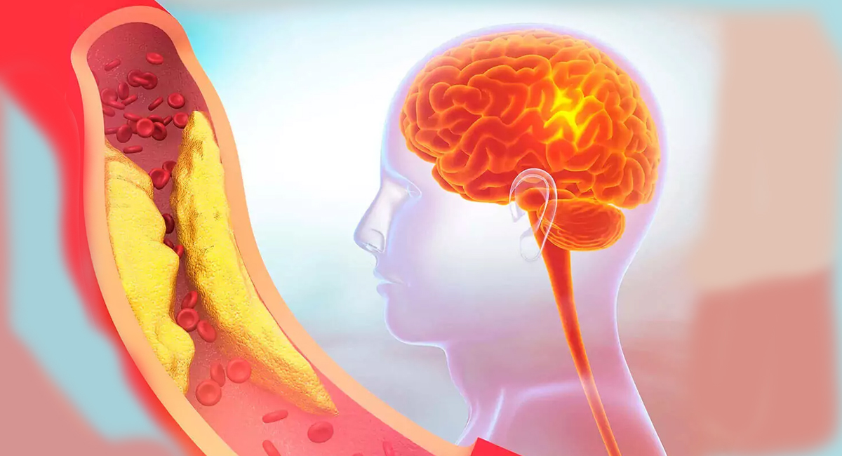
Arteries carry oxygenated blood away from the heart to the head and the body. There are two carotid arteries that supply blood to the brain. The carotid arteries lie on either side in the lower neck below the angle of the jaw. The carotid arteries supply blood to the front of the brain where thinking, speech, personality and sensory and motor functions reside. The vertebral arteries run through the spine and supply blood to the back part of the brain where brain stem and the cerebellum are present.
What is carotid artery disease?
Carotid artery disease is also called carotid artery stenosis which is due to atherosclerosis. Atherosclerosis is fat and other circulating substances (inflammatory cells, proteins, waste products, and calcium) deposition within the wall of the artery and called plaque. This plaque causes narrowing which progressively increases to block the artery and puts the patient at increased risk of stroke. Stroke is the 4th most common cause of death and the leading cause of permanent disability.
Who is prone to carotid artery disease?
These include:
- A family history of carotid artery disease, cardiac disease or peripheral artery disease.
- Smoking
- High blood pressure
- Diabetes
- Obesity
- Sedentary life
- Sleep apnea
- High cholesterol levels mainly LDL and triglycerides
Content Reviewed by - Dr. Jaisom Chopra
Symptoms
How do they present?
- The patient may be totally asymptomatic or there may be warning signs like mini-stroke lasting a few minutes to a few hours — Transient Ischemic Attack (TIA)
- Sudden loss of vision in one eye
- Sudden weakness or numbness on one side of the body or face.
- Sudden slurred speech making it difficult to understand
- Sudden loss of coordination
- Sudden dizziness or confusion
- Sudden difficulty in swallowing
- Sudden severe headache with unknown cause
When to see a doctor
Should you experience any of the above complaints go to the casualty because you are in danger of developing a full-fledged stroke.
What is a stroke?
This is also called 'brain attack' and occurs when the artery to the brain is blocked leading to the stoppage of blood flow and therefore lack of oxygen. Since the brain cannot store oxygen, therefore, it needs a constant supply of oxygenated blood and nutrients. When the blood supply is cut off for 3-4 minutes brain tissue begins to die leading to a stroke.
In which conditions will stroke occur?
- If the artery becomes narrowed or blocked by plaque.
- A blood clot travels to the artery and blocks it
- The artery in the brain bursts due to high blood pressure cutting the blood supply to the brain.
Diagnosis
How is carotid artery disease diagnosed?
- If you are at risk then it is advisable to have regular check-ups even if you are asymptomatic.
- A hissing sound when the doctor listens over the artery in the neck with a stethoscope strengthens the diagnosis
- Carotid Duplex Ultrasound - This is a non-invasive test and causes no inconvenience to the patient. It is fairly diagnostic in proving narrowing or blockage of the artery.
- Carotid Angiography - This is an invasive test where a catheter is put in the arm of the groin. A dye is pushed into the artery and the images recorded. It is conducted under local anesthesia and confirms narrowing or blockage after which further treatment can be decided stenting or surgery.
- Magnetic Resonance Angiography (MRA) - is a non-invasive magnetic scan with radio-waves providing information about the narrowing or the blockage of the arteries.
- CT Angiography - This gives three-dimensional pictures of the carotid and vertebral arteries, as well as the brain arteries.
How is carotid artery disease managed?
The three basic guidelines to manage this disease are:
- Making lifestyle changes
- Taking the prescribed drugs
- Having the procedure as recommended.
What are the lifestyle changes needed?
This prevents a carotid disease from progressing and they are:
- Quit smoking
- Control blood pressure, diabetes, heart disease and high cholesterol (LDL < 100 and HDL> 45)
- Regular check-ups by the doctors
- Diet control - low saturated fats, sodium, and cholesterol
- Weight control
- Daily 30 minutes of exercise
- Limit alcohol to less than 3 drinks per day.
- Manage other risk factors - patient with atrial fibrillation must have anticoagulation to prevent a clot forming and migrating to give rise to stroke.
What are the drugs indicated?
- Anti-platelets - reduce the risk of strokes and cardiac complications. They include Aspirin, Clopidogrel (prevent sticking of the platelets) and dipyridamole (Persantine). These could be given separately or in combination. At times anticoagulation like warfarin and acetone may be added to reduce the risk or stroke.
- Tissue Plasminogen activator (TPA) - It is a clot-dissolving drug used to treat stroke caused by blood clots (ischemic stroke). 80% of all strokes are ischemic. TPA only works if given within 3 hours of the start of stroke symptoms.
What are the corrective procedures?
The steps are as below:
The narrowed artery is dilated to increase blood flow to the brain. This dilatation is done by two methods
- Carotid stenting
- Carotid endarterectomy
- In both these procedures, the blood supply to the brain is brought to normal, which is exactly what is needed.
If however, the patient is asymptomatic despite the narrowing then one has to evaluate if medicines would be all that is needed or surgery would be better.
How is carotid endarterectomy performed?
- It is recommended for symptomatic patients with over 50% narrowing and asymptomatic patients with over 60% narrowing.
- It may be performed under general anesthesia or local anesthesia.
- An incision is made in the neck over the artery.
- The artery is exposed control taken.
- The plaque is removed after opening the artery.
- The artery is sewn and blood flow restored to the brain.
How is carotid angioplasty and stenting performed?
- The patient is awake.
- The catheter is inserted percutaneously in the groin or the arm under local anesthesia.
- The catheter is threaded towards the site of carotid stenosis under fluoroscopic guidance.
- A guide wire with a filter (umbrella) is placed beyond the narrowing on the side of the brain to prevent clots (debris) from going to the brain and causing stroke during the procedure. It is a protective device.
- A balloon is passed and dilated at the site of the narrowing to widen the narrow site.
- Now a metal stent or tube is placed at the widened site to prevent it from narrowing again.
- It takes several weeks for the inner artery lining to cover the stent from within.
- Studies have shown that carotid stenting with a protective device is as safe and effective in high-risk cases.
- The hospital stay in both cases is 2-3 days and people resume duty within one week.
- Do surgical patients need follow-up?
- This should be done at regular intervals.
How to find a doctor if you have a carotid disease?
The more complex the problem the more care you need and it is important you reach the right experts. Specialist medical care will have a direct impact on how well you do and how safe you are from having a stroke. It is recommended you contact a vascular surgery specialist.
Content Reviewed by - Dr. Jaisom Chopra